

Have you been vaccinated from the coronavirus? If so, you can credit the efficacy of your immunization to countless researchers. They tested numerous treatments on cell models, a necessary step in vaccine development. The most common model is a cell line, a specialized group of cells cultured for use in mimicking human responses. However, these cell lines usually grow in a flat, two-dimensional layer and may not be able to accurately model drug responses of a 3-D organ, such as the lungs. So, scientists used organoids as mini lungs for vaccine screening during the current pandemic.
Organoids are mini organ models, ranging in size from the width of a hair to five millimeters. As they grow, they naturally form into spheres. This system is unique because organoids are capable of self-renewal — dividing and forming new cells — and differentiation — changing from one cell type to another. These properties are necessary to accurately model a single cell’s interactions with its local environment, cell-to-cell interactions and physiological functions of the organ.
While lung organoids have benefited vaccine development for the novel coronavirus, they can also work their modeling magic on other diseases. Cancer researchers are starting to use organoids as they are useful and effective tools for testing anti-cancer compounds. A major reason why conventional cancer therapies fail is that cancers can vary greatly from patient to patient, meaning a drug that works in one patient may fail in another. Being able to quickly and accurately produce models from a patient’s cancer sample can provide researchers with the opportunity to screen and test drugs for that very same patient.
Dr. M. Laura Martin, Ex Vivo Models Director at the Weill Cornell Medicine Englander Institute for Precision Medicine has been working to establish organoid drug screening as a technology for individual patients. However, there are many challenges. For example, the microenvironment is extremely complex, containing “the extracellular matrix, the fibroblasts [connective tissue], the vascularity [blood flow, and] the immune cells,” and therefore it is difficult to model. To address this, Dr. Martin is using donor blood containing immune cells to grow alongside the tumor organoid and observe their interactions.
Another challenge is that some tumors are difficult to model in cell lines. For example, models for primary prostate cancer and breast cancer have extremely low rates of being successfully produced — at just 20-30%. Scientists are still trying to figure out how to sustain these types of tumors outside of the human body for study.
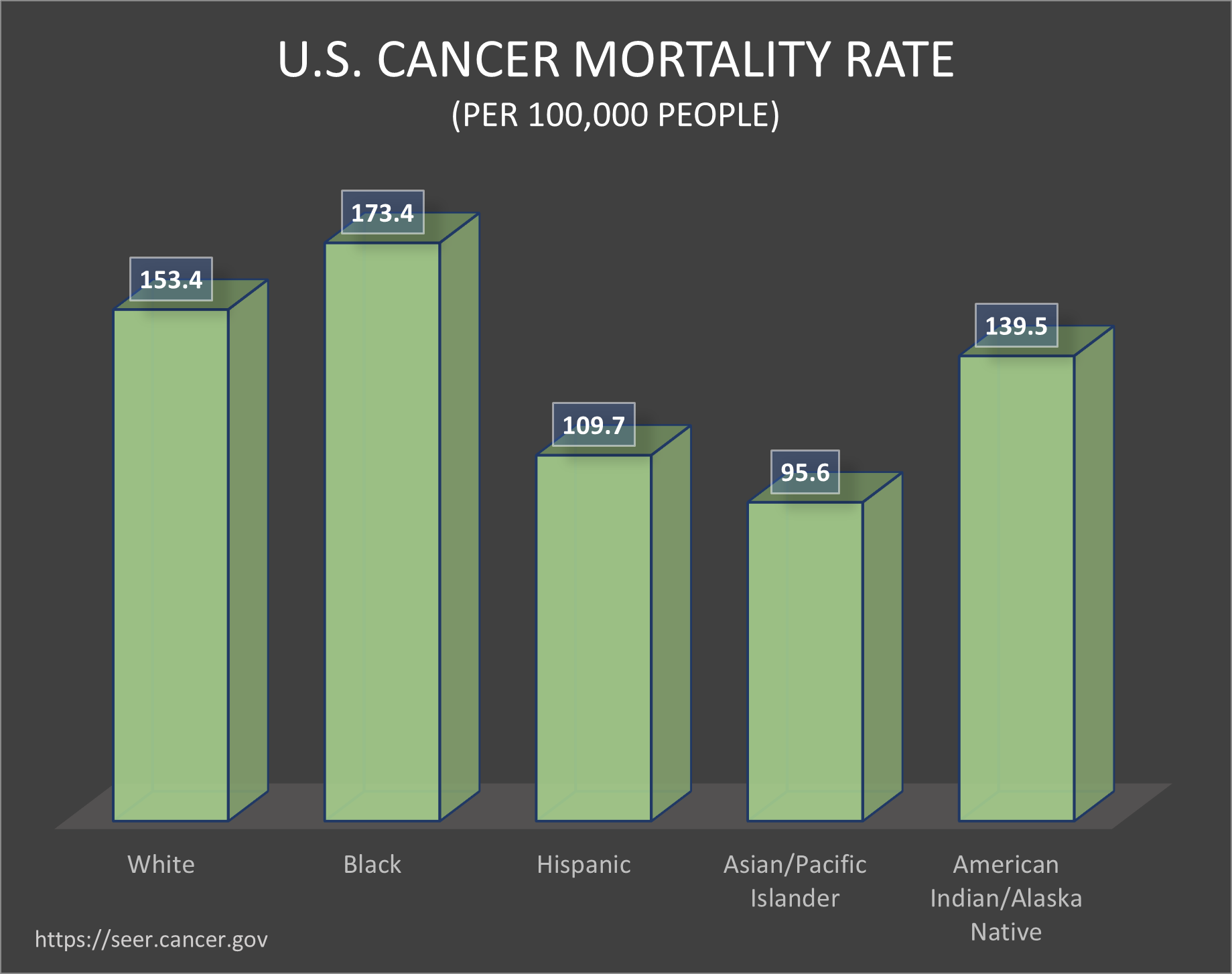
One area of cancer research that Dr. Martin is hoping to address through organoid technology is racial disparities in cancer treatments and outcomes. Within cancer, mortality and incidence rates impact people of different races unequally. Many modern drugs are designed to treat cancers with different mutations, which occur at varying rates across races. For example, Guerrero et al. reports Chinese, Korean and Japanese females with lung cancer had a higher prevalence of mutations in a gene called EGFR as compared to white females.
The lack of drugs available for cancer types occurring in people from racial and ethnic minority groups may be partially caused by a discrepancy in cell models. A vast majority of donor cells come from white patients. However, because organoids come from a patient’s own body, results derived from those organoids will be most applicable to them. This may mitigate underlying racial inequalities in treatment results.
At the same time, Dr. Martin and her colleagues are diversifying their cell samples so they more accurately reflect the general population. Together with Dr. Melissa B. Davis, an assistant professor of cell and developmental biology at Cornell, Dr. Martin is working in sub-Saharan countries in Africa, such as Ghana. Through the collaboration of many scientists, cell sourcings are becoming more and more varied, improving future models to reflect the types of cancers occurring for all people.
In the near future, we may be able to model individual cancer samples in organoids to create more personalized models for drug screening. If organoids are used in more patient trials and organoid research continues at the current pace, Dr. Martin hopes, in about five years, researchers will be able to identify a working drug for each individual patient.
- Organoids are three-dimensional research models created from stem cells. They are used to test cancer treatments. Their advantage compared to traditional 2-D cell lines is that they are better able to model organ systems.
- In this article, Dr. M. Laura Martin, Ex Vivo models director at the Englander Institute for Precision Medicine at Weill Cornell Medicine, discusses current challenges and potential improvements in the Ex Vivo models field.
- If the production of organoids can be scaled up, they may one day be used as interpatient models to test different cancer immunotherapies.
Sources
Barbuzano, Javier. “Organoids: A new window into disease, development and discovery.” Harvard Stem Cell Institute. 17 November 2017. https://hsci.harvard.edu/organoids
Drost, J., Clevers, H. “Organoids in cancer research.” Nat Rev Cancer 18, 407–418 (2018). https://doi.org/10.1038/s41568-018-0007-6
Fan, H., Demirci, U. & Chen, P. Emerging organoid models: leaping forward in cancer research. J Hematol Oncol 12, 142 (2019). https://doi.org/10.1186/s13045-019-0832-4
Guerrero et al. “Analysis of Racial/Ethnic Representation in Select Basic and Applied Cancer Research Studies.” doi: 10.1038/s41598-018-32264-x. 18 Sept 2018. https://www.ncbi.nlm.nih.gov/pmc/articles/PMC6143551/
Interview with Dr. Laura Martin. Interview by Rosie Liu. August 18, 2021.
Lindemans, Caroline A et al. “Interleukin-22 promotes intestinal-stem-cell-mediated epithelial regeneration.” Nature vol. 528,7583 (2015): 560-564. doi:10.1038/nature16460
Mallapaty, Smriti. “The mini lungs and other organoids helping to beat COVID.” Nature. 26 May 2021. https://www.nature.com/articles/d41586-021-01395-z
Yin, Xiaolei et al. “Engineering Stem Cell Organoids.” Cell stem cell vol. 18,1 (2016): 25-38. doi:10.1016/j.stem.2015.12.005
Editorial Team
- Chief Editor: Karishma Goswami
- Team Editor: Kevin Liu
- Creative Team Managers: Daniela Benoit, Bebe
Lemanowicz
- Social Media Team Manager: Spencer Lyudovyk
- Image Credits: Bebe Lemanowicz
Mentor
- Peggy Wang, Ph.D., is communications manager at the National Cancer Institute (NCI), where she works to inform researchers and the public about cancer genomics discoveries. She blogs, tweets and occasionally creates podcasts. Prior to joining the NCI, Peggy worked in bioinformatics to understand how DNA changes its shape and packaging to control gene expression. She earned her Ph.D. in biomedical engineering, imagining all the genes of the genome as a giant connected network.
Content Expert
M. Laura Martin, Ph.D., is the Ex Vivo Models Director at the Weill Cornell Medicine Englander Institute of Precision Medicine. She currently works on expanding Cornell’s ex-vivo models program and is hoping to use organoids to improve drug screening in patient care. She studied lipids which brought her to anti-cancer therapies. Excitement of finding new challenges in research helps her stay motivated.
