
In the Terminator movies, an artificial intelligence (AI) program named Skynet takes over the world. Skynet starts a nuclear war that destroys most of the human population and sends cyborgs known as Terminators to eliminate any survivors.
Whenever the topic of AI comes up, many people think of Skynet. However, this idea is a common misconception. AI isn’t one collective sentient being, but instead many separate computer systems that are independent of each other. AI systems perform tasks, such as identifying patterns in data, that once could only be accomplished with human intelligence. While AI is still developing, it has the potential to improve the world in many ways, especially in medicine.
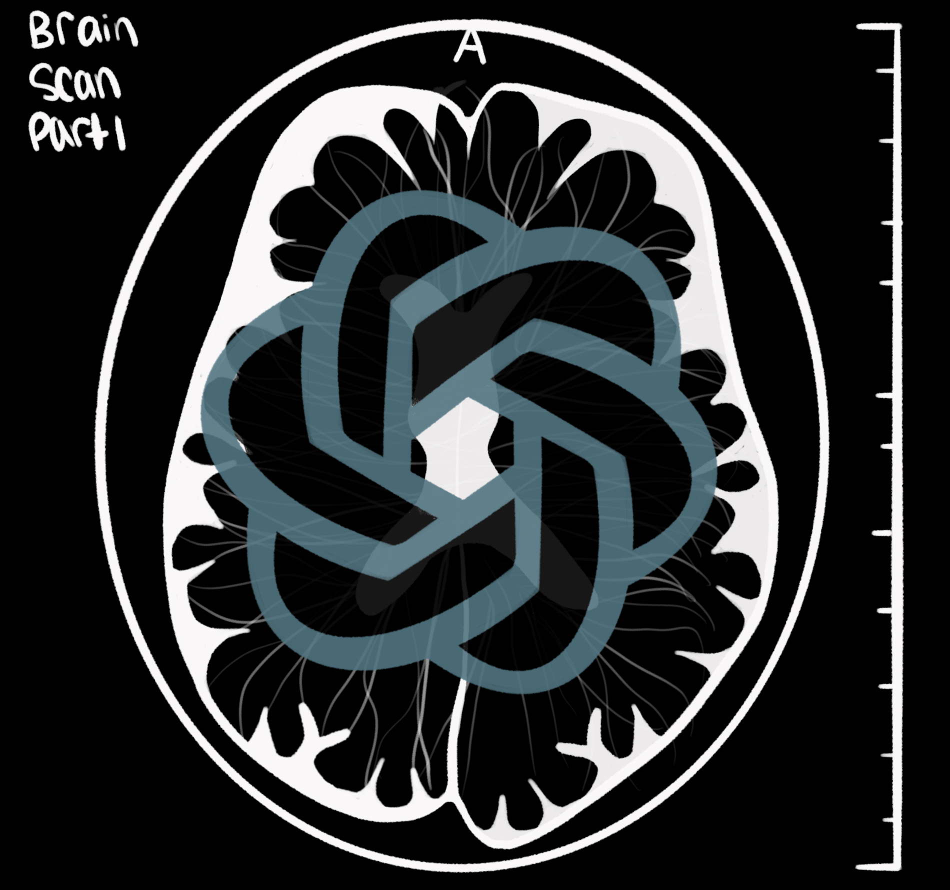
MRI, or magnetic resonance imaging, was developed in the 1970s as a groundbreaking medical tool. Imagine it as a high-tech camera that can see inside your body, but instead of using light, it uses powerful magnets and radio waves. Just as a detective uses different techniques to find hidden clues, MRI allows doctors to see detailed images of our organs and tissues, helping them diagnose various conditions and plan treatments more effectively. Compared to X-rays, which use harmful radiation, MRI is a safer way to look inside the entire body, from the brain to the joints.
Traditionally, MRI machines are large and heavy, taking up an entire room in a hospital. But now, scientists are developing smaller, portable MRI machines. While these new machines don’t produce images as clear as the bigger ones, they offer significant advantages. These smaller machines can be taken to remote places, such as small towns or villages far from hospitals. Smaller MRI machines are also cheaper and don’t need as much specialized equipment, making it easier for more people to get MRI scans, even if they don’t live near a big hospital. Using smaller machines, trades some picture quality for the ability to help more people in more places. These smaller machines could allow doctors to detect and treat health issues sooner in areas where they couldn’t before.
So, what does AI have to do with MRI imaging? AI, specifically, machine learning, can help improve the quality of these MRI images, making it easier for doctors to diagnose patients. Machine learning involves training AI on a lot of data to recognize trends and make predictions on new data it receives.
“AI is a sophisticated guessing tool. It basically looks for patterns,” says Andrew Dienstfrey, a researcher at NIST.
By analyzing patterns, AI can reproduce those patterns in the future when necessary. “Machine learning needs a lot of data to be able to recognize certain types of patterns,” he explains.
The idea of applying AI isn’t just a theoretical idea; researchers are testing the use of AI in the real world. In a 2023 study by NYU researchers, 170 participants received diagnostic knee scans.The researchers enhanced some of these MRI images with deep learning, while they left others unaltered. A group of radiologists, unaware of whether they were looking at enhanced or original images, evaluated the scans for ligament tears or other issues. The images were shown to the radiologists weeks apart to avoid memory bias. The study found that the AI-enhanced images made it easier for the radiologists to spot signs of injury.
While AI has made advancements, its future raises many ethical debates and uncertainties. Data privacy, specifically, is a major issue. These algorithms handle very sensitive medical data, and concerns arise about who can access this information and how it should be used.
Dr. Robert Barry of the NIBIB explains that there are certain solutions for protecting the privacy of individuals such as removing identifying features in certain images.
For example, in an MRI of someone’s whole head, “you can digitally change all the pixels from the eyes all the way around the ears and the jaw and change them to zeros,” he says. Solutions that creatively anonymize patient data ensure patients’ privacy and is something that researchers should keep in mind for the future.
We’re not facing Skynet, but rather a helping hand made of ones and zeros. As AI and MRI join forces, they’re painting a clearer picture of our insides, turning our bodies into less mysterious terrains. Even though there are still improvements to make, a combination of human brains and artificial smarts isn’t here to take over the world – it’s here to make our world healthier, one enhanced image at a time. So the next time you hear “AI,” don’t think of an apocalypse; think of it as a safer and healthier future.
- Scientists are developing smaller, portal MRI machines but this comes with decreased image quality.
- AI helps to look for patterns so that even lower quality images can provide helpful insights.
- AI can help doctors better identify abnormalities in MRI images regardless of the image quality.
Sources
Cargill, Michael. “Antigen Encounter and Presentation,” “B Cell Development and Function,” “T Cell Development and Function.” Immunology, North Carolina School of Science and Mathematics, Durham, North Carolina, April 2024.
DeRoo, J., Terry, J. S., Zhao, N., Stasevich, T. J., Snow, C. D., & Geiss, B. J. (2024). Pabfold: Linear antibody epitope prediction using AlphaFold2. eLife. https://doi.org/10.7554/elife.98369
JM, D., Grifoni, J, M., RR, G., Sette, & A, S. (2021, February 5). Sette Lab. La Jolla Institute for Immunology. https://www.lji.org/labs/sette/
Peters, Bjoern. “B Cell Epitope Prediction.” B Cell Epitope Prediction, 2020 User Workshop – 2.6, Immune Epitope Database and Analysis Resource, Virtual, November 5-6, 2020.
Ranganathan, S. (2019). Encyclopedia of Bioinformatics and Computational Biology. volume 1, methods. Elsevier.
Ras-Carmona, A., Lehmann, A. A., Lehmann, P. V., & Reche, P. A. (2022). Prediction of B cell epitopes in proteins using a novel sequence similarity-based method. Scientific Reports, 12(1). https://doi.org/10.1038/s41598-022-18021-1
Sanchez-Trincado, J. L., Gomez-Perosanz, M., & Reche, P. A. (2017). Fundamentals and methods for T- and B-cell epitope prediction. Journal of Immunology Research, 2017, 1–14. https://doi.org/10.1155/2017/2680160
Schaap-Johansen, A.-L., Vujović, M., Borch, A., Hadrup, S. R., & Marcatili, P. (2021). T cell epitope prediction and its application to immunotherapy. Frontiers in Immunology, 12. https://doi.org/10.3389/fimmu.2021.712488
Editorial Team
- Chief Editor: Katherine Mi
- Associate Editor: Christine Chen
- Team Editor: Riti Tandra
- Image Credit: Sylvia Xu
- Social Media Manager: Chloe Eng
- Social Media Team: Alexis Kim and Ellen Bu
Mentor
- Ben Stein, Managing Editor, Public Affairs Office, National Institute of Standards and Technology (NIST)
Content Expert
Andrew Dienstfrey, Ph.D. is a researcher at the National Institutes of Standards and Technology. He is an expert on the mathematics to computational physics and numerical analysis. His expertise in mathematics also applies to machine learning and artificial intelligence.
Robert Barry, Ph.D. is an expert in Biomedical Imaging and radiology. He has 20+ years of experience in MRI and has received a decade of continuous NIBIB funding to develop novel hardware and acquisition methodologies for non-invasive functional imaging of the human spinal cord. His expertise in the past several years has grown to the field of AI reconstruction of MRI images.