You’re a scientist in a lab, and you’ve worked tirelessly to develop a treatment for a disease. After a decade’s worth of research, your drug has finally passed animal screening and is on its way to a clinical trial. While you may be towards the end of the drug development pathway, the finish line still remains far out of reach. In fact, 90% of drugs fail clinical trials despite success in earlier phases of drug development, effectively wasting years of researcher efforts and perhaps billions of dollars. The research and pharmaceutical industry needs to do something differently to raise this success rate. Though biochemistry comes to mind when thinking of pharmaceuticals, some researchers have turned to engineering for a potential solution. Ever heard of bioprinting?
Bioprinting — the 3D printing of tissue from live cells — includes a broad range of technology, some even comparable to the traditional inkjet printing used in the office. Bioprinting utilizes an ink, known as a bioink, to form the tissue. The bioink is typically composed of cells, which gives the tissue its functionality, as well as polymers such as alginate or gelatin that can support cellular life by providing structure and organization. Ultimately, the bioprinted construct should resemble tissue found in an animal, such as a liver or bladder, providing researchers with a realistic model to test for drug efficacy and toxicity.
“each broken heart [and] each broken bone is different from person-to-person,”
Dr. Shaochen Chen
Dr. Shaochen Chen, a professor at the University of California, San Diego and a leading researcher in the field, has worked on a variety of projects, ranging from printing cardiac tissue to tumor models. An engineer at heart, Dr. Chen explains that it’s “in my DNA to solve problems,” highlighting his engineering background as a contributor to his success.
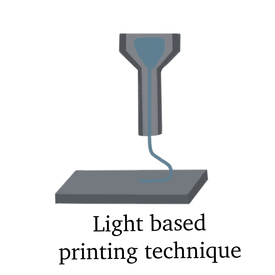
While there are many different methods of bioprinting, Dr. Chen utilizes a light-based technique. Remember that blue light used to solidify your fillings at the dentist? The process with this form of bioprinting is similar. Rather than fillings, researchers take a liquid bioink and expose the polymer to UV light, which will then harden the solution. The printer follows a pattern provided by a digital design, and the tissue is formed layer-by-layer, a technique known as additive manufacturing. Once the tissue is printed in well plates, researchers can administer drugs to the model and observe the effects. Toxicity can be predicted prior to human, or even animal, exposure.

One advantage of bioprinting is its versatility, as a variety of miniature organs have already been constructed. Dr. Chen explains that his lab has worked on heart, liver, and brain tissue, amongst that of other vital organs. Additionally, researchers have also focused on disease modeling, or printing diseased tissue, which can provide scientists with accurate models that they can use to find treatments or even cures. While cancer models have been a main focus, Dr. Chen believes that bioprinting may be an important technology in the development of new treatments for a variety of diseases.
Bioprinting opens the door to personalized medicine, the idea of creating a treatment specific to a single patient by first creating models directly from that patient’s cells. As Dr. Chen explains, “each broken heart [and] each broken bone is different from person-to-person,” meaning a treatment that works for one patient may not work for another. This technology can help doctors find treatments best suited to specific patients, which could become a reality within the next 10-15 years, as speculated by Dr. Chen. Other applications include tissue printed for wound healing, such as nerve repair or skin grafting, as well as pairings with other bioengineering methods, such as organ-on-chip research, or devices with microchannels that allow for the culture of miniature tissues. Lastly, engineers are cautiously optimistic that full-size organs may one day be printed, a hope that could change the lives of over 100,000 people waiting for transplants.
Let’s travel back to the start of our story: you’re a scientist in a lab, and you’ve worked tirelessly to develop a treatment for a disease. But this time, you have a bioprinted liver model at your disposal. After testing your product on the tissue, you see evidence of drug-induced toxicity. While it may initially be disappointing, this model just saved both animals and patients from harm, not to mention millions of dollars and countless hours of work, as this toxic effect was caught much earlier in the process. Though it may seem small, bioprinting truly does have the potential to not only improve lives, but maybe even save some.
- Bioprinting uses 3D printing of live cells to create tissue models for drug testing, helping detect toxicity early and potentially reducing the high failure rate of clinical trials.
- Bioprinting offers a new way to test drugs using 3D-printed tissues that mimic human organs, improving drug safety and reducing costs. Dr. Shaochen Chen’s work highlights its potential in personalized medicine and the eventual printing of full-size organs.
Sources
- Extrusion vs. DLP 3D bioprinting – explanatory comparison. CELLINK. (2023, June 15). https://www.cellink.com/blog/extrusion-vs-dlp-3d-bioprinting-explanatory-comparison/
- Mesko, B. (2022, June 1). 3D bioprinting – overview of how bioprinting will break into healthcare. The Medical Futurist. https://medicalfuturist.com/3d-bioprinting-overview/ Organ donation statistics. (n.d.). https://www.organdonor.gov/learn/organ-donation-statistics
- Persaud, A., Maus, A., Strait, L., & Zhu, D. (2022). 3D bioprinting with live cells. Engineered Regeneration, 3(3), 292–309. https://doi.org/10.1016/j.engreg.2022.07.002
- Rosen, E. (2020, July 27). A possible weapon against the pandemic: Printing human tissue. The New York Times. https://www.nytimes.com/2020/07/27/science/bioprinting-covid-19-tests.html
- Sun, D., Gao, W., Hu, H., & Zhou, S. (2022). Why 90% of clinical drug development fails and how to improve it? Acta Pharmaceutica Sinica B, 12(7), 3049–3062. https://doi.org/10.1016/j.apsb.2022.02.002
- Tabatabaei Rezaei, N., Kumar, H., Liu, H., Lee, S. S., Park, S. S., & Kim, K. (2023). Recent advances in organ‐on‐chips integrated with Bioprinting Technologies for Drug Screening. Advanced Healthcare Materials, 12(20). https://doi.org/10.1002/adhm.202203172
- X, S. (2023, June 12). Taking biofabrication to the next level: Innovations in volumetric bioprinting. Phys.org. https://phys.org/news/2023-06-biofabrication-volumetric-bioprinting.html
- Yan, J., Li, Z., Guo, J., Liu, S., & Guo, J. (2022). Organ-on-a-chip: A new tool for in vitro research. Biosensors and Bioelectronics, 216, 114626. https://doi.org/10.1016/j.bios.2022.114626
- You, S., Xiang, Y., Hwang, H. H., Berry, D. B., Kiratitanaporn, W., Guan, J., Yao, E., Tang, M., Zhong, Z., Ma, X., Wangpraseurt, D., Sun, Y., Lu, T., & Chen, S. (2023). High cell density and high-resolution 3D bioprinting for fabricating vascularized tissues. Science Advances, 9(8). https://doi.org/10.1126/sciadv.ade7923
Editorial Team
- Chief Editor: Annika Singh
- Team Editor: Tara Prakash
- Image Credit: Sylvia Xu
- Social Media Lead: Chloe Eng
Mentor
- Peggy Wang is the communications manager at the National Cancer Institute (NCI), where she works to inform researchers and the public about cancer genomics discoveries. She blogs, tweets, and occasionally creates podcasts. Prior to NCI, Peggy worked in bioinformatics to understand how DNA changes its shape and packaging to control gene expression. She earned her Ph.D. in biomedical engineering, imagining all the genes of the genome as a giant connected network.
Content Expert
Shaochen Chen, Ph.D. is a professor of nanoengineering at the University of California, San Diego. Dr. Chen’s lab has focused its research on bioprinting, tissue engineering, biomaterials and nanomaterials, as well as organ-on-chip applications.

